
When an optical biometer fails to read through a dense cataract, clinics need a reliable fallback. That’s exactly where DGH A steps in. This compact ultrasound biometry device gives eye care professionals accurate axial length measurements, anterior chamber depth, and lens thickness data — all critical inputs for intraocular lens power calculation before cataract surgery.
- What Is DGH A?
- Why DGH A Is Important
- How DGH A Works
- A Guide to Ocular Biometry and the Importance of DGH A-Scan Ultrasound
- Applanation Versus Immersion: Choosing the Right Technique with DGH A
- Standout Features of the Portable DGH A Ultrasound Device
- Applications of DGH A
- Applications in Ophthalmology and Eye Care
- Application of DGH A-Scan in Government and Public Healthcare Institutions
- Use of DGH A-Scan in Corporate and Professional Environments
- DGH A Scan Accuracy: How It Compares to Optical Biometry and Other Devices
- Practical Guide: How to Use DGH A for IOL Power Calculation in Cataract Surgery
- Benefits of DGH A
- Challenges Associated With DGH A
- Best Practices for Using DGH A
- Essential Maintenance: Cleaning and Care of DGH A Scan Probes
- DGH A-Scan Integration in Clinics: Enhancing Workflow and Software Performance, Workflow and Software Benefits
- Future Relevance and Advancements of DGH A
- Conclusion
- FAQs
- What is DGH A and how does it work?
- What is the difference between applanation and immersion technique in DGH A?
- How does the accuracy of DGH A-Scan compare to optical biometry?
- Does DGH A support post-refractive IOL calculations?
- How do you clean and maintain DGH A scan probes?
- Is DGH compatible with clinic EMR and software systems?
- What eye conditions can DGH A help diagnose?
- What is the full form and medical meaning of DGH?
Unlike many tools that promise precision but add workflow complexity, the DGH A is straightforward, portable, and clinically trusted. Whether you’re planning a surgical case or tracking myopia progression in a young patient, this device covers both scenarios without requiring a cart, a console, or a complicated setup.
This guide covers everything from how the device works to how it fits into a modern clinic — with practical detail at every step.
What Is DGH A?
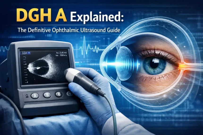
The DGH A is a portable ophthalmic ultrasound device designed for ocular biometry. It is also sold under the name Scanmate 6000 and connects to any standard Windows computer via USB. The probe weighs under a pound, which makes it easy to move between exam rooms or even transport to satellite clinics.
At its core, DGH A uses a 10 MHz ultrasonic transducer to send sound waves through the eye and capture returning echoes from the cornea, lens surfaces, and retina. These echoes generate precise measurements used in IOL calculation formulas.
Meaning, Definition, and Purpose of DGH A
The term DGH A refers to a specific model of diagnostic ultrasound device produced for ophthalmic use. Its purpose is standardization of biometric measurement — giving clinicians a consistent, reproducible reference point for surgical planning.
Within a clinical framework, DGH A fills a defined role: it provides the axial length measurement and additional biometric data needed when optical devices cannot deliver a reliable reading. That role doesn’t change across practice sizes. A high-volume surgical center and a small general practice both use it in the same way.
History and Development of DGH A Technology
Development of DGH A technology began in the early 2000s, when clinicians identified a gap between available imaging tools and what surgical precision demanded. Early prototypes focused on image processing clarity and minimizing invasive steps during examinations.
By 2010, the technology had matured into a frontrunner in ophthalmic ultrasound, guided by feedback from clinicians who needed better user interfaces and faster workflows. Collaborations with tech companies pushed real-time analysis features into the product. The result was a device that balanced clinical precision with everyday usability — without requiring extensive training to operate.
Why DGH A Is Important
Accurate biometry drives successful surgical outcomes. A measurement error of just 0.1 mm in axial length can shift refraction by nearly a full diopter. For patients receiving premium IOLs, that margin matters enormously.
Importance of Administrative and Regulatory Systems
Beyond the operating room, DGH A also functions as a classification reference in structured clinical and regulatory environments. Administrative settings rely on clearly defined measurement standards to reduce misclassification, avoid delays, and ensure procedures follow established compliance requirements. Regulatory bodies often tie operational approval categories to documented biometry protocols, which is where a consistent tool like DGH A adds institutional value.
Role of DGH A in Decision-Making and Governance
Decision-makers in surgical planning use DGH A data as a core reference point. When two measurement methods produce different results, the immersion ultrasound reading often serves as the tiebreaker. Clear data reduces ambiguity in approvals and IOL selection. It also reduces errors caused by operator variability — a significant concern in high-volume settings where multiple technicians handle biometry.
How DGH A Works
The device captures biometric data by firing sound waves through the eye and timing the returning echoes. Different tissue layers — cornea, anterior lens surface, posterior lens surface, retina — each return a distinct echo spike. Software maps these spikes into precise measurements.
Structural Framework of DGH A
DGH A operates within a tiered measurement system. Each scan is automatically graded using a three-star pattern recognition system built into the Scanmate software. A three-star result signals high-quality waveform alignment. Lower ratings prompt the technician to reposition and rescan. This hierarchy prevents borderline measurements from slipping through unreviewed.
The software also supports automatic gain control, which adjusts waveform amplification in real time so the technician doesn’t have to manually tune settings for each patient.
Criteria for Assigning and Validating DGH A Measurements
Valid measurements require consistent probe alignment, correct velocity settings for the patient’s lens status (phakic, pseudophakic, or aphakic), and a minimum of five to ten reproducible scans. The software flags outliers and allows manual waveform review when results fall outside acceptable repeatability thresholds.
Periodic calibration and validation against known phantoms maintain system integrity over time — a requirement in most clinical quality frameworks.
A Guide to Ocular Biometry and the Importance of DGH A-Scan Ultrasound
Ocular biometry measures the physical dimensions of the eye to calculate the correct IOL power before surgery. Axial length sits at the center of every IOL formula — from SRK/T to Barrett Universal II. Sound waves from the 10 MHz ultrasonic transducer travel through the cornea, aqueous humor, lens, and vitreous before hitting the retina, producing echo data that maps each layer’s position.
Where optical biometers rely on light and fail in eyes with dense cataracts, small pupils, or significant corneal irregularities, ultrasound continues to function. DGH A’s pattern recognition software evaluates each waveform instantly and generates axial length progression reports useful for myopia management — tracking changes in growing eyes over months and years.
Applanation Versus Immersion: Choosing the Right Technique with DGH A
Both techniques are available with DGH A, and each has a clear clinical use case.
Contact (Applanation) Mode
- Faster and simpler for routine cases
- Probe touches the anesthetized cornea directly
- Patient fixates on the red light built into the probe tip
- The compression lockout feature prevents artificially short readings from corneal indentation
Immersion Mode
- Uses the included Prager shell filled with sterile saline
- The shell rests on the sclera; the probe is suspended in the fluid bath
- No corneal contact means zero compression artifact
- Repeatability typically within ±0.03 mm
- Preferred for premium IOL patients and post-refractive cases
Most experienced surgeons default to immersion whenever maximum precision matters. The audible feedback and real-time waveform display help guide probe alignment in both modes.
Standout Features of the Portable DGH A Ultrasound Device
| Feature | Detail |
| Device name | Scanmate 6000 |
| Connectivity | USB to Windows PC |
| Weight | Under 1 lb |
| Transducer frequency | 10 MHz |
| Scan grading | Automatic three-star system |
| Gain control | Automatic |
| IOL formulas | Multiple built-in + post-refractive options |
| EMR compatibility | Yes — direct export |
| Myopia reports | Axial length progression tracking |
| Manual review | Available for borderline scans |
The Scanmate software stores patient details, runs IOL calculations, and exports reports to your EMR in a single workflow. There’s no separate console, no waiting for a system to boot, and no tech support required for daily operation.
Applications of DGH A
Applications in Ophthalmology and Eye Care
DGH A’s primary clinical role is cataract surgery planning, but its application list extends further:
- Diabetic retinopathy screening — high-resolution imaging aids early detection
- Glaucoma progression monitoring — axial length changes tracked over time
- Age-related macular degeneration (AMD) — cross-sectional data support severity assessment
- Preoperative planning — corneal topography review and biometric mapping before surgery
- Myopia management — axial length progression reports for pediatric patients
For any condition where retinal structures, anterior chamber depth, or lens status need quantification, DGH A provides usable data even when optical approaches fail.
Application of DGH A-Scan in Government and Public Healthcare Institutions
In government healthcare systems and public institutions, DGH A supports operational-level compliance by providing a standardized biometric measurement tool that meets regulatory classifications. Uniform implementation of documented biometry protocols is often tied to institutional approval categories and funding criteria.
Use of DGH A-Scan in Corporate and Professional Environments
Private practices and corporate ophthalmology groups use DGH A to align clinical processes with certification standards and internal grading systems. It supports staff compliance training and ensures departments meet organizational objectives around surgical outcomes and documentation accuracy.
DGH A Scan Accuracy: How It Compares to Optical Biometry and Other Devices
| Method | Best For | Limitation |
| Optical biometry | Clear media, non-contact | Fails with dense cataracts |
| DGH A applanation | Quick routine scans | Risk of corneal compression |
| DGH A immersion | Premium IOL, post-refractive | Slightly more setup time |
| Fundus photography | Retinal imaging | No biometric data |
| OCT | Structural retinal detail | Limited axial length data |
Studies and clinical practice consistently show that DGH A immersion technique matches optical biometry results closely in clear-media eyes. In eyes with media opacities, DGH A often provides the only usable data. Industry estimates suggest optical biometers fail in 8 to 17 percent of cataract cases — exactly where ultrasound earns its place.
Practical Guide: How to Use DGH A for IOL Power Calculation in Cataract Surgery
- Open the Scanmate software and create or load the patient record
- Select contact or immersion mode based on clinical indication
- Set the correct velocity for the patient’s lens status (phakic, pseudophakic, aphakic)
- Apply a topical anesthetic; prepare saline for immersion if needed
- Position the probe, instruct fixation, and acquire five to ten consistent scans
- Review waveforms — software auto-selects best measurements, or you review manually
- Switch to the IOL calculator, choose your preferred formula, and compare lens models
- Export the finalized report to your EMR
The entire workflow typically takes under five minutes for a cooperative patient.
Benefits of DGH A
- Clarity — standardized measurement removes ambiguity from surgical planning
- Portability — moves between exam rooms, satellite clinics, and mobile units without hassle
- Efficiency — compact design and intuitive software reduce time per patient
- Fairness — consistent criteria ensure equal measurement quality across technicians
- Flexibility — supports home visits and mobile clinics without sacrificing diagnostic quality
- Compatibility — EMR export and EHR integration work with most existing practice systems
Challenges Associated With DGH A
No tool is without limits. Applanation mode carries a risk of corneal compression if the technique is poor, producing artificially short axial length readings. In very large systems managing multiple classification categories alongside DGH A data, complexity can increase without proper documentation and staff training.
Misinterpretation of scan quality grades is another common issue, particularly with newer technicians who haven’t yet developed confidence reading waveform patterns. Regular updates to velocity settings and formula selections are also necessary as new IOL models enter the market.
Best Practices for Using DGH A
- Provide clear documentation on scan grading standards for all staff
- Run structured training programs focused on immersion technique before technicians handle premium IOL cases
- Schedule periodic evaluation of measurement consistency — compare contact vs. immersion results on the same eye
- Add probe cleaning to the end-of-day checklist to maintain infection control compliance
- Review axial length progression reports quarterly for myopia management patients
Small procedural habits — consistent fixation instruction, correct velocity selection, methodical waveform review — produce the tightest repeatability and the most reliable IOL outcomes.
Essential Maintenance: Cleaning and Care of DGH A Scan Probes
After every patient contact, disconnect the probe and clean the tip, housing, and cord with a soft cloth dampened in mild soap and water, or 70 percent isopropyl alcohol. Follow immediately with an approved disinfectant per manufacturer guidelines.
Never submerge the probe beyond the strain relief point. Dry it fully before storage. The immersion shell follows the same protocol. A daily inspection of the cable and USB connection prevents unexpected downtime — a small habit that keeps the device reliable for years.
DGH A-Scan Integration in Clinics: Enhancing Workflow and Software Performance, Workflow and Software Benefits
Because DGH A runs on the same Windows computer used for charting, it fits naturally into existing workflows. Patient data flows directly into the practice database. Reports print or export with a single click, and EHR systems receive data without manual re-entry.
High-volume surgical centers benefit from the personalized lens constants feature inside Scanmate software. After a few dozen cases, surgeons refine their constants — tightening refractive outcomes over time. Smaller practices gain from the portability: one unit can rotate across multiple exam rooms without disrupting scheduling.
Future Relevance and Advancements of DGH A
The next phase of DGH A development points toward artificial intelligence integration. AI-assisted pattern recognition could identify measurement outliers faster than current software and flag cases where additional review is warranted. Miniaturization may produce even lighter portable versions suited for underserved areas and remote telemedicine deployments.
As digital platforms increasingly handle automated processing and tracking of clinical data, DGH A’s EMR compatibility positions it well for integration into broader digital health frameworks. Regulatory demands around documentation and compliance will likely drive adoption in markets currently relying on manual biometric records.
Conclusion
DGH A occupies a specific and important role in ophthalmic care — delivering reliable biometric data when optical systems cannot. Its combination of portability, proven accuracy, and Scanmate software integration makes it a practical tool for clinics of any size. Beyond eye care, its function as a standardized classification reference reflects a broader truth: structured, consistent measurement tools reduce errors and improve outcomes across every domain where precision matters. For surgeons, technicians, and administrators alike, understanding DGH A means being better prepared for the cases that demand a reliable fallback.
FAQs
What is DGH A and how does it work?
DGH A is a portable ophthalmic ultrasound device that measures the eye’s internal dimensions using a 10 MHz transducer. It captures axial length, anterior chamber depth, and lens thickness data — all essential for IOL calculation before cataract surgery. The device is non-invasive, provides real-time feedback, and connects to a Windows PC via USB.
What is the difference between applanation and immersion technique in DGH A?
Applanation places the probe directly on the anesthetized cornea and is faster for routine scans. Immersion uses a saline-filled Prager shell resting on the sclera, eliminating corneal compression. Immersion delivers tighter repeatability (often ±0.03 mm) and is the preferred method for premium IOL patients.
How does the accuracy of DGH A-Scan compare to optical biometry?
In eyes with clear media, optical biometry leads slightly due to its non-contact nature. For eyes with dense cataracts or other media opacities — roughly 8 to 17 percent of cases — DGH A immersion technique often provides the only usable biometric data, matching optical results closely when proper technique is applied.
Does DGH A support post-refractive IOL calculations?
Yes. The Scanmate software includes built-in post-refractive formulas for patients who previously had LASIK, PRK, or RK. Surgeons can calculate lens power confidently using these tools without relying on external calculators.
How do you clean and maintain DGH A scan probes?
Wipe the probe tip, housing, and cord with 70 percent isopropyl alcohol or mild soap after each use, followed by an approved disinfectant per manufacturer guidelines. Never submerge the probe past the strain relief. Apply the same protocol to the immersion shell, and perform a daily USB connection and cable inspection.
Is DGH compatible with clinic EMR and software systems?
Yes. DGH A runs on standard Windows PCs and exports data directly to EHR and EMR systems. This allows seamless data sharing across healthcare teams without manual re-entry, making it compatible with most existing clinic software environments.
What eye conditions can DGH A help diagnose?
DGH A supports the diagnosis and monitoring of diabetic retinopathy, glaucoma, cataracts, age-related macular degeneration (AMD), and retinal diseases. It also tracks axial length progression for myopia management and assists in preoperative planning for patients with corneal irregularities.
What is the full form and medical meaning of DGH?
DGH has several meanings depending on context. In medicine, it stands for Diploma in Global Health, awarded by the Royal College of Physicians with MSF. A DGH hospital refers to a District General Hospital serving local and rural populations. In India, DGH stands for the Directorate General of Hydrocarbons under the Ministry of Petroleum & Natural Gas. In water chemistry, dGH measures water hardness based on calcium and magnesium carbonate levels per liter.